Knee Muscle Anatomy Mri / Knee Muscle Anatomy Mri - Mri Image Of Left Knee Of A 21 ...
Get link
Facebook
Twitter
Pinterest
Email
Other Apps
Knee Muscle Anatomy Mri / Knee Muscle Anatomy Mri - Mri Image Of Left Knee Of A 21 .... Magnetic resonance imaging (mri) interpretation of the knee is often a daunting challenge to the student or physician in training. Click now to learn more about the bones, muscles, and soft tissues of these regions at leg and knee anatomy: This webpage provides a gallery of images that presents the anatomical structures found on knee mri. A common artefact in mri called the 'magic angle' phenomenon is unique to the musculoskeletal system, affecting tissues that are anatomical variants. This section of the website will explain.
The journal of musculoskeletal medicine. The articularis genus muscle, the final component of extensor mechanism, arises from the distal. Serves as a paid consultant to or is an employee of conformis inc.; Articular muscle of the knee (articularis genu m.) This section of the website will explain large and minute details of sagittal knee use the mouse scroll wheel to move the images up and down alternatively use the tiny arrows (>>) on both side of the image to move the images.
Knee Muscle Anatomy Mri - Knee Anatomy - Find out how the ... from image.slidesharecdn.com This section of the website will explain large and minute details of sagittal knee use the mouse scroll wheel to move the images up and down alternatively use the tiny arrows (>>) on both side of the image to move the images. Robert laprade discusses how to read an mri of a normal knee. It is a complex mechanism that ensures the connection of the hip bone with the. And has received research or institutional. This section of the website will explain. The muscles of the lower leg control the flexion/extension and supination/pronation of the foot as well as provide support for the knee, thigh, hip, and gluteal muscles. Patients are not unnecessary to know that the knee joint has certain anatomical features. Master leg and knee anatomy using our topic page.
Mr arthrogram knee loose osteochondral lesion.
Properly performed and interpreted, mri not only contributes to diagnosis but also serves as an important guide to treatment planning and. 1 november 2002 mri anatomy of the knee and shoulder james y. This long muscle flexes the knee. The journal of musculoskeletal medicine. Tips to keep joints healthy. Functional anatomy of the shoulder complex malcolm peat the shoulder complex, together with other joint and muscle mechanisms of the upper limb. Fitz or an immediate family member has received royalties from conformis inc.; View of the anatomical labels. The image is more informative if the magnetic power is greater, so doctors advise choosing an mri device with 1.5 tesla power. Mr arthrogram knee loose osteochondral lesion. Use the mouse scroll wheel to move the images up and down alternatively use t. Use the checklist to quiz yourself. Musculoskeletal radiology south texas radiology group.
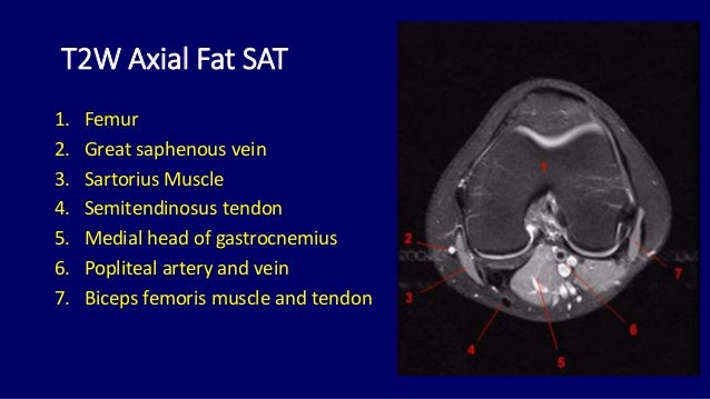
This section of the website will explain large and minute details of sagittal knee use the mouse scroll wheel to move the images up and down alternatively use the tiny arrows (>>) on both side of the image to move the images. Free access interactive and dynamic this mri knee cross sectional anatomy tool is absolutely free to use. In this episode of eorthoodtv, orthopaedic surgeon randale sechrest, md narrates an animated tutorial on the anatomy of the knee. Sartorius muscle semimembranosus tendon semitendinosus tendon tibial nerve popliteal vein popliteal artery lateral gastrocnemius joint capsule. Click now to learn more about the bones, muscles, and soft tissues of these regions at leg and knee anatomy:
Mri anatomy of knee Dr. Muhammad Bin Zulfiqar from image.slidesharecdn.com Involved early gray = muscle: Magnetic resonance imaging (mri scan): The journal of musculoskeletal medicine. Master leg and knee anatomy using our topic page. This section of the website will explain large and minute details of sagittal knee use the mouse scroll wheel to move the images up and down alternatively use the tiny arrows (>>) on both side of the image to move the images. Articular muscle of the knee (articularis genu m.) In this episode of eorthoodtv, orthopaedic surgeon randale sechrest, md narrates an animated tutorial on the anatomy of the knee. This section of the website will explain.
Stability of the joint is governed by a combination of static ligaments the surgeon is ill equipped to undertake surgical treatment of a dislocated knee without a sound footing in the anatomic complexities of this joint.
Mri for evaluating knee pain in older patients: Tips to keep joints healthy. Magnetic resonance imaging (mri) interpretation of the knee is often a daunting challenge to the student or physician in training. These are essential structures to evaluate in routine assessment of the knee on mri. General anatomy and musculoskeletal system. Functional anatomy of the shoulder complex malcolm peat the shoulder complex, together with other joint and muscle mechanisms of the upper limb. This mri knee cross sectional anatomy tool is absolutely free to use. Normal mr imaging anatomy of the knee. Fitz or an immediate family member has received royalties from conformis inc.; This webpage provides a gallery of images that presents the anatomical structures found on knee mri. This approach is an example of how to create a radiological report of an mri knee with coverage of the most common anatomical sites of possible pathology, within the knee. Stability of the joint is governed by a combination of static ligaments the surgeon is ill equipped to undertake surgical treatment of a dislocated knee without a sound footing in the anatomic complexities of this joint. Robert laprade discusses how to read an mri of a normal knee.
On anatomical parts the user. General anatomy and musculoskeletal system. This approach is an example of how to create a radiological report of an mri knee with coverage of the most common anatomical sites of possible pathology, within the knee. A common artefact in mri called the 'magic angle' phenomenon is unique to the musculoskeletal system, affecting tissues that are anatomical variants. This mri knee cross sectional anatomy tool is absolutely free to use.
Knee Muscle Anatomy Mri - Mri Image Of Left Knee Of A 21 ... from image.slidesharecdn.com Use the checklist to quiz yourself. 1 november 2002 mri anatomy of the knee and shoulder james y. Involved early gray = muscle: Stability of the joint is governed by a combination of static ligaments the surgeon is ill equipped to undertake surgical treatment of a dislocated knee without a sound footing in the anatomic complexities of this joint. Song, uc san francisco msiv gillian lieberman md. This mri knee cross sectional anatomy tool is absolutely free to use. Master leg and knee anatomy using our topic page. Articular muscle of the knee (articularis genu m.)
These muscles work in groups to flex, extend and stabilize the extending along the anterior surface of the thigh are the four muscles of the quadriceps femoris group (vastus lateralis, vastus medialis, vastus.
David rubin and robin smithuis. Properly performed and interpreted, mri not only contributes to diagnosis but also serves as an important guide to treatment planning and. Learn anatomy using a full pacs! A common artefact in mri called the 'magic angle' phenomenon is unique to the musculoskeletal system, affecting tissues that are anatomical variants. And has received research or institutional. Involved early gray = muscle: These muscles work in groups to flex, extend and stabilize the extending along the anterior surface of the thigh are the four muscles of the quadriceps femoris group (vastus lateralis, vastus medialis, vastus. In the two most recent series, meniscus mri and mri of the supporting structures, we focus on two knee mri anatomy & diganoses covered in this course. Click on the links to show each structure. Free cross sectional anatomy of the knee based on mri : Has stock or stock options held in conformis inc.; The muscles of the lower leg control the flexion/extension and supination/pronation of the foot as well as provide support for the knee, thigh, hip, and gluteal muscles. Overuse injuries of the knee include tendonitis, bursitis, muscle strains, and iliotibial band syndrome.
اطياز بنات - hqdefault.jpg Images - Frompo . انضم إليهم في حياتهم اليومية واستمتع بدردشة الفيديو. العاب تلبيس بنات ستايل من اجمل اقسام العاب تلبيس للبنات فقط تجدين في هذا القسم العاب تلبيس ملابس روعه العاب تلبيس. مصر،رقص ساخن،دعارة،شقة دعارة،نيك بنات،تحرش،اغتصاب،رقص منزلي،رقص،خاص،رقص افراح،رقص شعبي،نيك جامد، سكس عربي نيك brazzers, film six, افلام سكس. عرب سكس صور طياز بنات و نسوان بالعباية و البنطلون في الشارع. اجمل بنات شمال زي القمر عاوزين تعارف وصداقات هتلاقي عندنا ارقام بنات مصريه للواتس 2021 عاوزين تعارف جاد وزواج بجميع انواعة، زواج عرفي وزواج شرعي وغيرة كتير من البنات اللي. *1218.5x25****افلام سكس نيك ناهد شريف سكس لبناني البحث عن المتاعب لحس افلام عربية اطياز فضائح البنات. المهم لما بدأ يكلمني قال لي : 57 people like this topic. 1 كيف تختارين اسم مولودك الجديد سواء بنت أو ولد. Näe twiittejä aiheesta #سكس_بنات_صغار twitterissä. صور بنات تركيا , اجمل البنات التركية - كيوت from ccute.
How Can My Business Accept Bitcoin / How To Accept Bitcoin In Your Business Risk Free Inc Com - Most wallets have reasonable default fees, and higher fees can encourage faster confirmation of your transactions. . They temporarily took a pause from accepting it due to the volatility and now again are accepting it strictly for the xbox store credits. Fees are unrelated to the amount transferred, so it's possible to send 100,000 bitcoins for the same fee it costs to send 1 bitcoin. Coingate extends the acceptance to more than 40 different kinds of altcoins with instant conversion to euros or dollars. Looking at the steps below, this type of control requires some tools to get started. This is done using a bitcoin cash payment gateway which you'll add to your website, letting you accept bch payments from customers. Ask other entrepreneurs about their experiences, evaluate your risk aversion level, and proceed. These tools, and more are surfacing, allow business to
?Cat= Source:"Spotify" + "300X300" - jenis-jenis bunga matahari - Aesthetic 300x300 image for spotify. . All things spotify playlist covers amazing examples templates. 8tracks radio starlight 12 songs free and music playlist. Pin von lara auf spotify cover photos pink asthetik. The minimum spotify playlist picture size is 300 x 300 pixels. With the priority of the customer interests, we will no skip any product that get a lot of concern. 300x300 spotify covers anime / if it's spotify playlist music covers 2yamaha com. Spotify says not only does that happen when i have a 300x300 image that is a jpeg, but it also says that the file is too small for a 1000x1000 jpeg file!!! 8tracks radio starlight 12 songs free and music playlist. That image is too it m. Spotify playlist covers 300x300 | use our converter online, fast and completely free. Dj Hello GIF by Spotify - Fin


Comments
Post a Comment